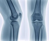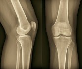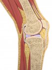
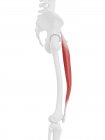
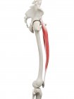
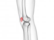
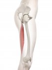
Knee joint anatomy — Stock photos
ID: 160225312
By:ScienceRF
"Knee joint anatomy" is an authentic stock image by ScienceRF. It’s available in the following resolutions: 1600 x 1600px, 2600 x 2600px, 4180 x 4180px. The minimum price for an image is 49$. Image in the highest quality is 4180 x 4180px, 300 dpi, and costs 449$.
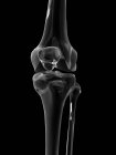
Similar Images


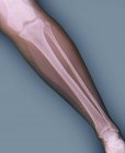
Same Series