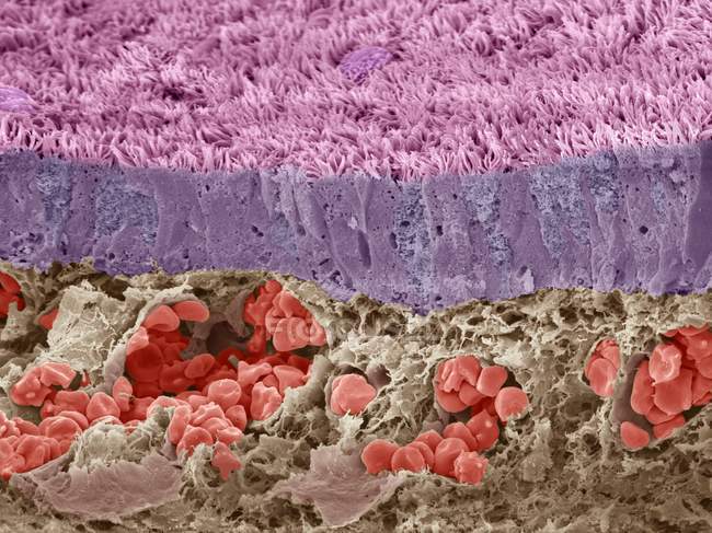
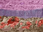
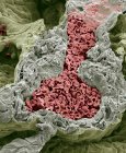
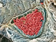
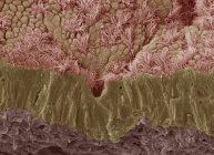
Coloured scanning electron micrograph (SEM) of a section through a vein in the liver, which is filled with red blood cells (erythrocytes, red). — Stock photos
ID: 160558438
By:ScienceRF
"Coloured scanning electron micrograph (SEM) of a section through a vein in the liver, which is filled with red blood cells (erythrocytes, red)." is an authentic stock image by ScienceRF. It’s available in the following resolutions: 1600 x 1199px, 2600 x 1948px, 4886 x 3661px. The minimum price for an image is 49$. Image in the highest quality is 4886 x 3661px, 300 dpi, and costs 449$.
Similar Images


Same Series