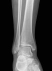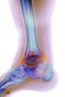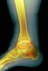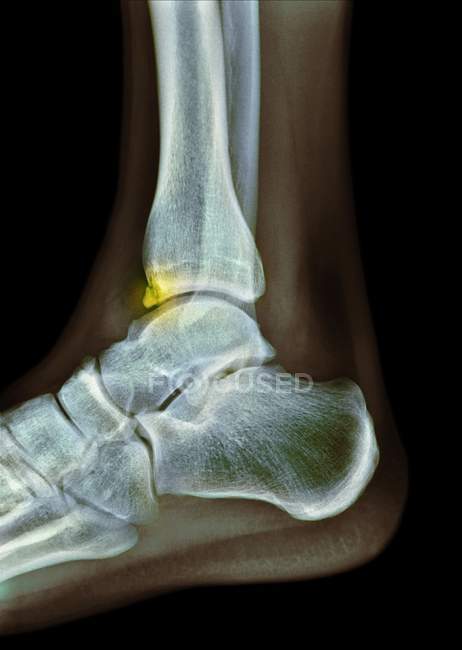
Coloured X-ray of the foot of a 22-year-old male patient with a spur (osteophyte, highlighted) affecting the tibia (shin bone). — Stock photos
ID: 160563166
By:ScienceRF
"Coloured X-ray of the foot of a 22-year-old male patient with a spur (osteophyte, highlighted) affecting the tibia (shin bone)." is an authentic stock image by ScienceRF. It’s available in the following resolutions: 1138 x 1600px, 1849 x 2600px, 3570 x 5020px. The minimum price for an image is 49$. Image in the highest quality is 3570 x 5020px, 300 dpi, and costs 449$.
Similar Images