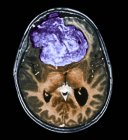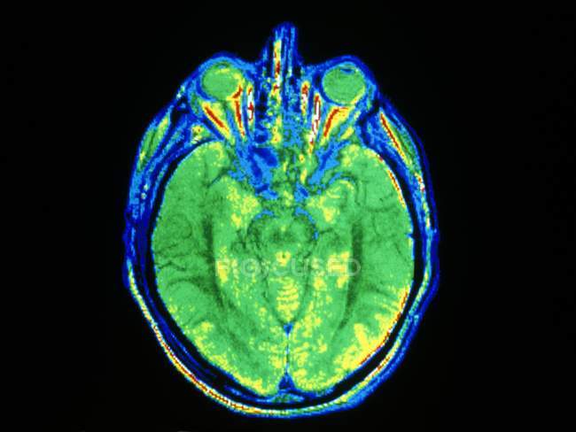
False-colour Magnetic Resonance Image (MRI) of an axial section through a human head, showing the division of the bulk of the brain into left and right cerebral hemispheres. — Stock photos
ID: 160568102
By:ScienceRF
"False-colour Magnetic Resonance Image (MRI) of an axial section through a human head, showing the division of the bulk of the brain into left and right cerebral hemispheres." is an authentic stock image by ScienceRF. It’s available in the following resolutions: 1600 x 1202px, 2600 x 1954px, 4903 x 3684px. The minimum price for an image is 49$. Image in the highest quality is 4903 x 3684px, 300 dpi, and costs 449$.
Similar Images