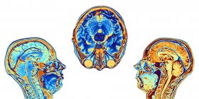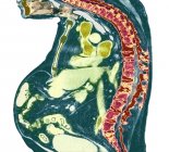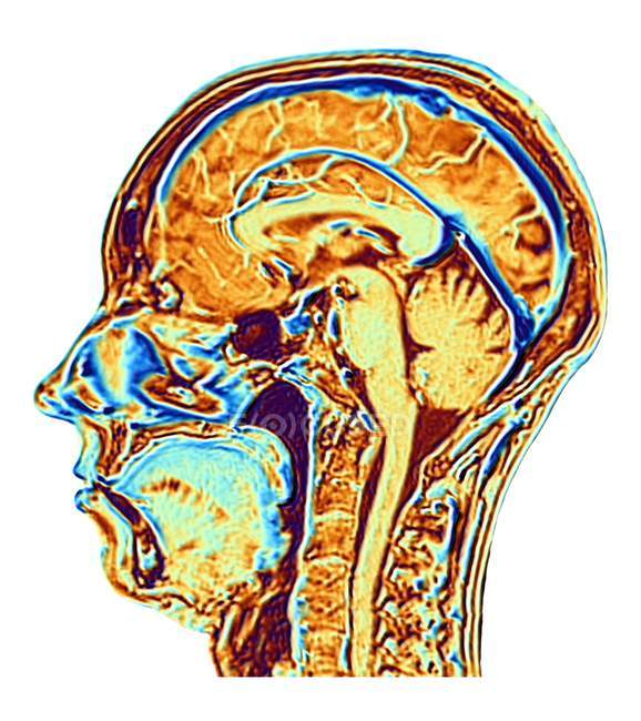
Computer enhanced false-colour Magnetic Resonance Image (MRI) of a mid-sagittal section through the head of a normal 46 year-old woman, showing structures of the brain, spine and facial tissues. — Stock photos
ID: 161674320
By:ScienceRF
"Computer enhanced false-colour Magnetic Resonance Image (MRI) of a mid-sagittal section through the head of a normal 46 year-old woman, showing structures of the brain, spine and facial tissues." is an authentic stock image by ScienceRF. It’s available in the following resolutions: 1422 x 1600px, 2311 x 2600px, 4800 x 5400px. The minimum price for an image is 49$. Image in the highest quality is 4800 x 5400px, 300 dpi, and costs 449$.
Similar Images