En
  • En English
  • De Deutsch
  • Fr Français
  • Sp Español
  • Ru Русский
  • It Italiano
  • Pt Português
  • Ua Українська
Sign In Sign Up
Sign In Sign Up
    Dividing prostate gland cancer cells, colored scanning electron micrograph. — Stock Photo

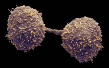
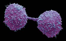
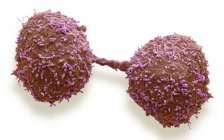
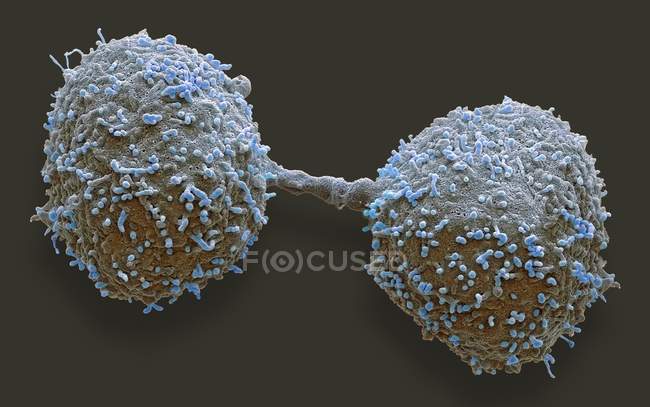
    Dividing prostate gland cancer cells, colored scanning electron micrograph. — Stock photos

    ID:  318068216
    By:ScienceRF
    "Dividing prostate gland cancer cells, colored scanning electron micrograph." is an authentic stock image by ScienceRF. It’s available in the following resolutions: 1600 x 1002px, 2600 x 1628px, 5284 x 3308px. The minimum price for an image is 49$. Image in the highest quality is 5284 x 3308px, 300 dpi, and costs 449$.

    Similar Images

    Colored scanning electron micrograph of cancer cells from human colon. — Stock Photo
    Colored scanning electron micrograph of cancer cells from human colon. — Stock Photo
    White blood cells — Stock Photo
    Asian sweets on plate — Stock Photo
    Entamoeba histolytica engulfing red blood cells — Stock Photo
    Coloured scanning electron micrograph of 293T cell infected with human immunodeficiency virus. — Stock Photo
    Primrose (Primula sp.) pollen grain, coloured scanning electron micrograph (SEM). — Stock Photo
    Colored scanning electron micrograph of human red blood cells erythrocytes, white blood cells leukocytes and platelet thrombocyte. — Stock Photo
    Coloured scanning electron micrograph (SEM) of human myeloid white blood cells found in bone marrow. — Stock Photo
    Puffball fungus spore — Stock Photo
    Fresh partly peeled Lychee — Stock Photo
    Pralines with dried fruits — Stock Photo
    3d illustration of brain cancer cell connecting to tissue. — Stock Photo
    Chocolate truffles on plate — Stock Photo
    Lymphocytes white blood cells, digital illustration. — Stock Photo
    Puffball fungus spore — Stock Photo
    Black truffles, whole and halved — Stock Photo

    Same Series

    Dividing prostate gland cancer cells, colored scanning electron micrograph. — Stock Photo
    Dividing prostate gland cancer cells, colored scanning electron micrograph. — Stock Photo
    Dividing prostate gland cancer cells, colored scanning electron micrograph. — Stock Photo

    Keywords

    • Cancer
    • 3d
    • Pathological
    • Malignant
    • Cells
    • Micrograph
    • Medicine
    • Histopathological
    • 3d rendering
    • Science
    • Sem
    • Medical
    • Disorder
    • Prostate gland
    • Tumor
    • Microscopic
    • Anatomy
    • Body
    • Prostate cancer
    • Growth
    • Human anatomy
    • Disease
    • Prostatic
    • Histopathology
    • Illustration
    • Dividing
    • Scanning electron micrograph
    • Digital
    • Cancer cells
    • Colored
    • False coloured
    • Anatomical
    • Oncology
    • Prostate
    • Malignancy
    • Pathology
    • Tumour

    Company

    About Press New Stock Images

    Legal Information

    Terms of Use License Agreement Privacy Policy

    Contact

    +1-954-990-0075 Contact Us

    Follow us

    © 2025 Focused ® Premium Stock Photos. All rights reserved.