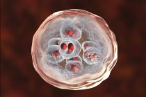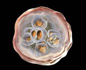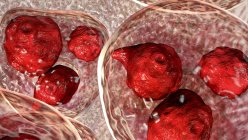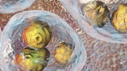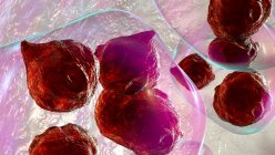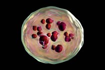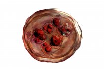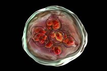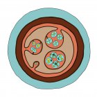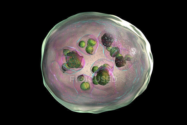
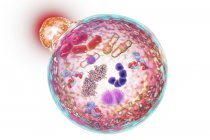
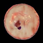
Echinococcus granulosus hydatid cyst, computer illustration — Stock photos
ID: 360110460
By:ScienceRF
"Echinococcus granulosus hydatid cyst, computer illustration" is an authentic stock image by ScienceRF. It’s available in the following resolutions: 1600 x 1067px, 2600 x 1733px, 6556 x 4370px. The minimum price for an image is 49$. Image in the highest quality is 6556 x 4370px, 300 dpi, and costs 449$.
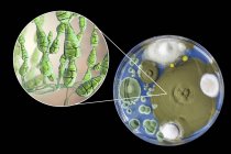
Similar Images
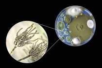


Same Series