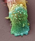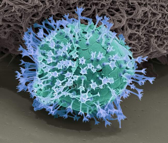
Acanthocystis. Coloured scanning electron micrograph (SEM) of a centrohelid heliozoan with tangential plate-scales and radial spines — Stock photos
ID: 379645002
By:ScienceRF
"Acanthocystis. Coloured scanning electron micrograph (SEM) of a centrohelid heliozoan with tangential plate-scales and radial spines" is an authentic stock image by ScienceRF. It’s available in the following resolutions: 1600 x 1366px, 2600 x 2219px, 4572 x 3902px. The minimum price for an image is 49$. Image in the highest quality is 4572 x 3902px, 300 dpi, and costs 449$.
Similar Images