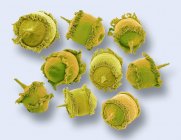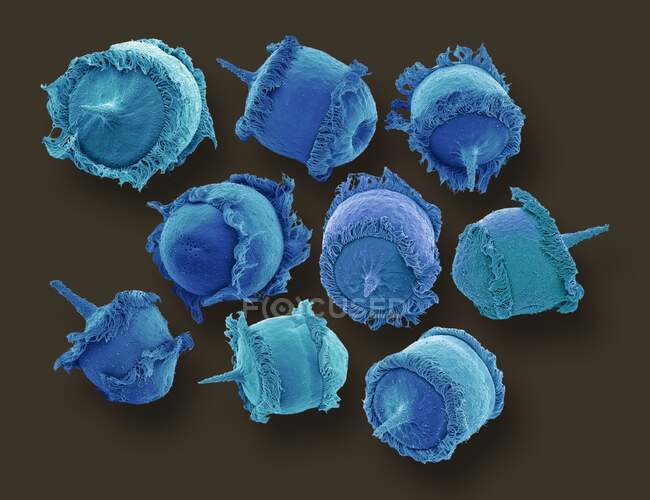
Didinium sp. ciliate protozoa, coloured scanning electron micrograph (SEM). These tiny single-celled organisms are found in freshwater and marine habitats. They are predatory organism, feeding on other ciliate protozoans, mainly Paramecium — Stock photos
ID: 399647656
By:ScienceRF
"Didinium sp. ciliate protozoa, coloured scanning electron micrograph (SEM). These tiny single-celled organisms are found in freshwater and marine habitats. They are predatory organism, feeding on other ciliate protozoans, mainly Paramecium" is an authentic stock image by ScienceRF. It’s available in the following resolutions: 1600 x 1231px, 2600 x 2000px, 4953 x 3810px. The minimum price for an image is 49$. Image in the highest quality is 4953 x 3810px, 300 dpi, and costs 449$.
Similar Images


Same Series