
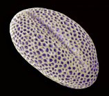
Maize root. Light micrograph (LM) of section through root of maize plant (Zea mays) showing typical monocot arrangement of vascular bundles. central vascular cylinder is comprised of central cluster of parenchyma cells — Stock photos
ID: 441641290
By:ScienceRF
"Maize root. Light micrograph (LM) of section through root of maize plant (Zea mays) showing typical monocot arrangement of vascular bundles. central vascular cylinder is comprised of central cluster of parenchyma cells" is an authentic stock image by ScienceRF. It’s available in the following resolutions: 1555 x 1600px, 2527 x 2600px, 4444 x 4572px. The minimum price for an image is 49$. Image in the highest quality is 4444 x 4572px, 300 dpi, and costs 449$.
Similar Images


Same Series

