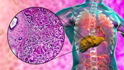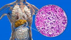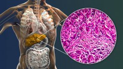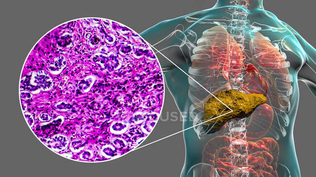
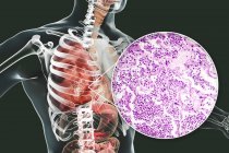
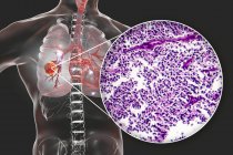
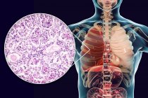
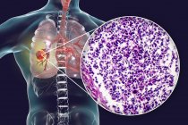
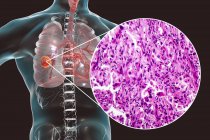
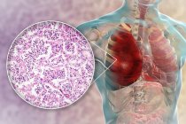
Liver cirrhosis. Computer illustration and light micrograph of a section through a human liver with from primary biliary cirrhosis. Cirrhosis is a disease in which bands of fibrosis (internal scarring) break up the internal structure of the liver — Stock photos
ID: 450152876
By:ScienceRF
"Liver cirrhosis. Computer illustration and light micrograph of a section through a human liver with from primary biliary cirrhosis. Cirrhosis is a disease in which bands of fibrosis (internal scarring) break up the internal structure of the liver" is an authentic stock image by ScienceRF. It’s available in the following resolutions: 1600 x 900px, 2600 x 1462px, 5570 x 3133px. The minimum price for an image is 49$. Image in the highest quality is 5570 x 3133px, 300 dpi, and costs 449$.
Similar Images


Same Series