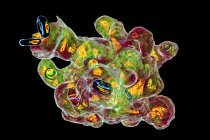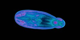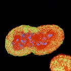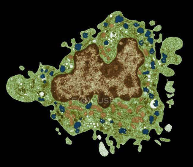
Macrophage. Coloured transmission electron micrograph (TEM) of a macrophage cell. The cell's nucleus is brown. Mitochondria (orange) in the cell's cytoplasm produce energy for the cell. Lysosomes (blue) contain enzymes for digesting foreign particles — Stock photos
ID: 463649568
By:ScienceRF
"Macrophage. Coloured transmission electron micrograph (TEM) of a macrophage cell. The cell's nucleus is brown. Mitochondria (orange) in the cell's cytoplasm produce energy for the cell. Lysosomes (blue) contain enzymes for digesting foreign particles" is an authentic stock image by ScienceRF. It’s available in the following resolutions: 1600 x 1385px, 2600 x 2251px, 4572 x 3958px. The minimum price for an image is 49$. Image in the highest quality is 4572 x 3958px, 300 dpi, and costs 449$.
Similar Images