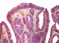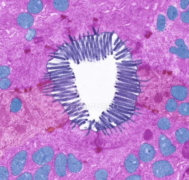
Intestinal microvilli. Coloured transmission electron micrograph (TEM) of a section through microvilli from the small intestine. These tiny structures (cyan) form a dense brush-like covering on the absorptive surfaces of the cells — Stock photos
ID: 463649596
By:ScienceRF
"Intestinal microvilli. Coloured transmission electron micrograph (TEM) of a section through microvilli from the small intestine. These tiny structures (cyan) form a dense brush-like covering on the absorptive surfaces of the cells" is an authentic stock image by ScienceRF. It’s available in the following resolutions: 1600 x 1524px, 2600 x 2477px, 4572 x 4356px. The minimum price for an image is 49$. Image in the highest quality is 4572 x 4356px, 300 dpi, and costs 449$.
Similar Images