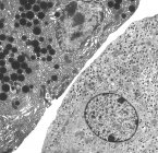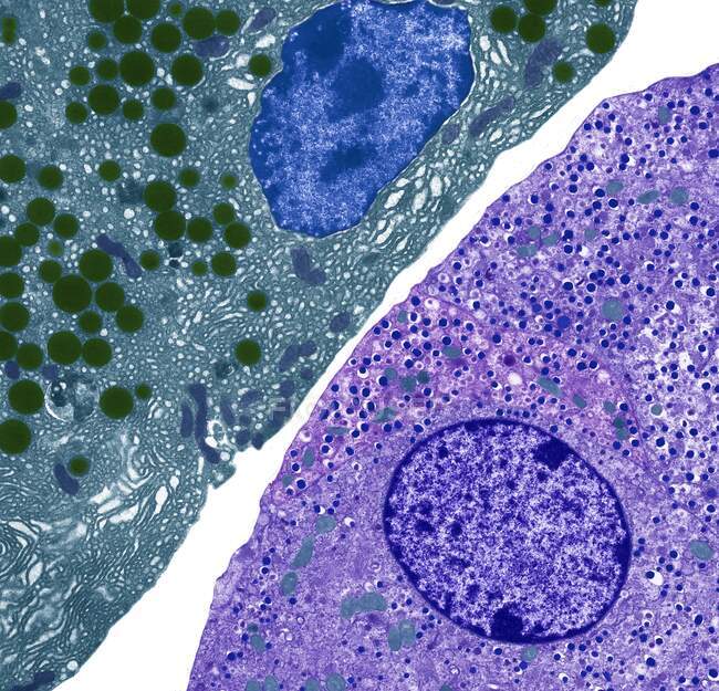
Pancreatic cells. Colored transmission electron micrograph (TEM) of acinar (exocrine) pancreatic cells (red) adjacent to hormone- secreting (endocrine) Islet of Langerhans cells (yellow) — Stock photos
ID: 463649636
By:ScienceRF
"Pancreatic cells. Colored transmission electron micrograph (TEM) of acinar (exocrine) pancreatic cells (red) adjacent to hormone- secreting (endocrine) Islet of Langerhans cells (yellow)" is an authentic stock image by ScienceRF. It’s available in the following resolutions: 1600 x 1539px, 2600 x 2501px, 4572 x 4398px. The minimum price for an image is 49$. Image in the highest quality is 4572 x 4398px, 300 dpi, and costs 449$.
Similar Images


Same Series