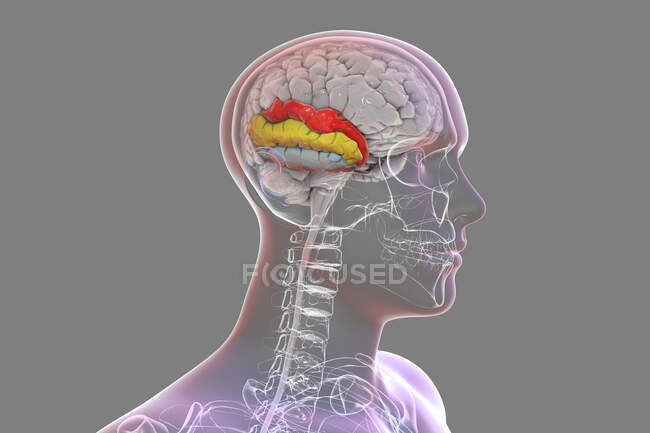
Human brain with highlighted temporal gyri, computer illustration. This is showing the superior temporal (red), middle (yellow), and inferior (blue) gyri. They are involved in processing auditory information and encoding of memory. — Stock photos
ID: 470161184
By:ScienceRF
"Human brain with highlighted temporal gyri, computer illustration. This is showing the superior temporal (red), middle (yellow), and inferior (blue) gyri. They are involved in processing auditory information and encoding of memory." is an authentic stock image by ScienceRF. It’s available in the following resolutions: 1600 x 1067px, 2600 x 1733px, 6000 x 4000px. The minimum price for an image is 49$. Image in the highest quality is 6000 x 4000px, 300 dpi, and costs 449$.
Similar Images