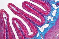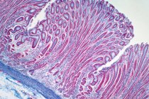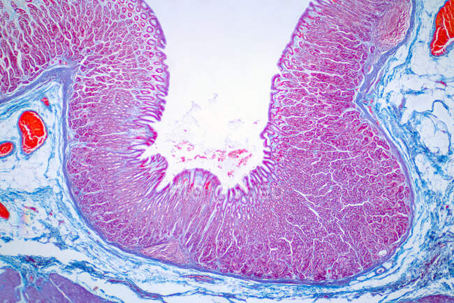
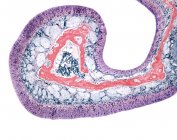
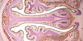
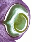
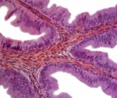
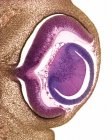
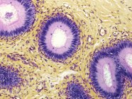
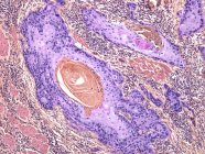
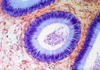
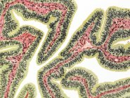
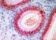
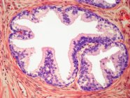
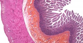
Light micrograph of a colon biopsy from a colonoscopy. The pathology report describes normal colonic mucosa fragment with colic glands. Haematoxylin and eosin stain. — Stock photos
ID: 494266462
By:ScienceRF
"Light micrograph of a colon biopsy from a colonoscopy. The pathology report describes normal colonic mucosa fragment with colic glands. Haematoxylin and eosin stain." is an authentic stock image by ScienceRF. It’s available in the following resolutions: 1600 x 1067px, 2600 x 1734px, 8192 x 5464px. The minimum price for an image is 49$. Image in the highest quality is 8192 x 5464px, 300 dpi, and costs 449$.
Similar Images
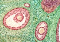
Same Series