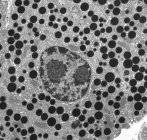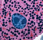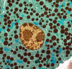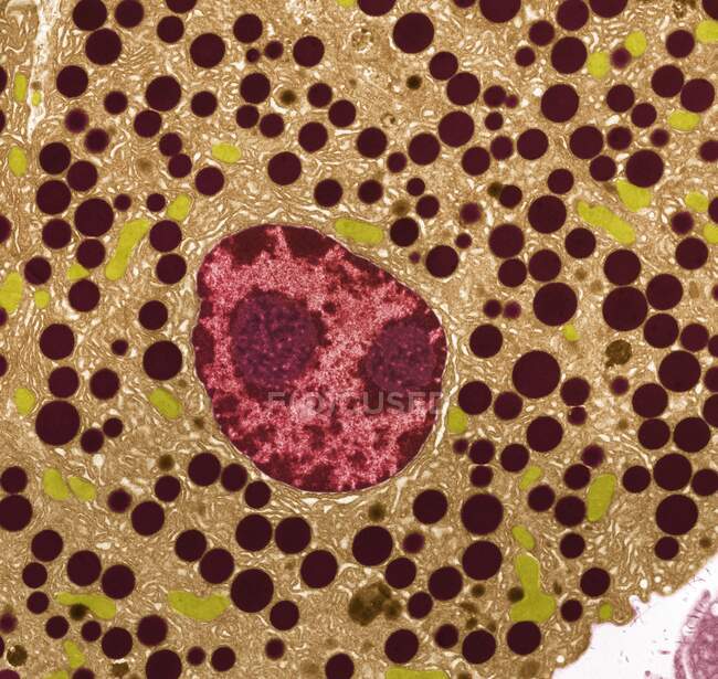
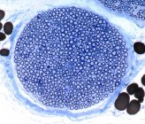
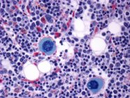
Pancreas tissue. Transmission electron micrograph (TEM) of part of the exocrine pancreas. Seen here are zymogen granules and cell nuclei. In the image the endoplasmic reticulum that fills the cytoplasm is clearly visible. — Stock photos
ID: 463649590
By:ScienceRF
"Pancreas tissue. Transmission electron micrograph (TEM) of part of the exocrine pancreas. Seen here are zymogen granules and cell nuclei. In the image the endoplasmic reticulum that fills the cytoplasm is clearly visible." is an authentic stock image by ScienceRF. It’s available in the following resolutions: 1600 x 1515px, 2600 x 2462px, 4572 x 4329px. The minimum price for an image is 49$. Image in the highest quality is 4572 x 4329px, 300 dpi, and costs 449$.
Similar Images


Same Series