En
  • En English
  • De Deutsch
  • Fr Français
  • Sp Español
  • Ru Русский
  • It Italiano
  • Pt Português
  • Ua Українська
Sign In Sign Up
Sign In Sign Up
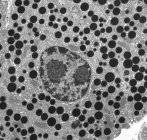
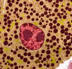
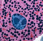
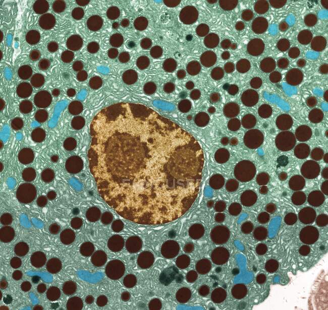
    Pancreas tissue. Transmission electron micrograph (TEM) of part of the exocrine pancreas. Seen here are zymogen granules and cell nuclei. In the image the endoplasmic reticulum that fills the cytoplasm is clearly visible. — Stock Photo

    Pancreas tissue. Transmission electron micrograph (TEM) of part of the exocrine pancreas. Seen here are zymogen granules and cell nuclei. In the image the endoplasmic reticulum that fills the cytoplasm is clearly visible. — Stock photos

    ID:  463649616
    By:ScienceRF
    "Pancreas tissue. Transmission electron micrograph (TEM) of part of the exocrine pancreas. Seen here are zymogen granules and cell nuclei. In the image the endoplasmic reticulum that fills the cytoplasm is clearly visible." is an authentic stock image by ScienceRF. It’s available in the following resolutions: 1600 x 1515px, 2600 x 2462px, 4572 x 4329px. The minimum price for an image is 49$. Image in the highest quality is 4572 x 4329px, 300 dpi, and costs 449$.

    Similar Images

    Directly above view of bowls filled with cereals wheat flakes. — Stock Photo
    Testicular cancer or teratoma — Stock Photo
    Directly above view of bowls filled with buckwheat and other cereals wheat flakes. — Stock Photo
    Section through bone marrow — Stock Photo
    Hepatitis C and Polio virus particles, digital illustration. — Stock Photo
    Blueberry pie on polka-dot napkin — Stock Photo
    Section through bone marrow — Stock Photo
    Coxsackievirus enterovirus virus particles, digital illustration. — Stock Photo
    Seminiferous tubules and lending cells — Stock Photo
    Polio virus particles, digital illustration. — Stock Photo
    Colorful cups of coffee on table, high angle view. — Stock Photo
    Making filter coffee — Stock Photo
    Directly above view of ceramic bowls filled with cereals and wheat flakes. — Stock Photo
    Nerve bundle from the sciatic nerve — Stock Photo
    Top view of red lentils in bowl and on cloth — Stock Photo
    Making filter coffee. — Stock Photo
    Coloured scanning electron micrograph (SEM) of a freeze-fractured water lily stem showing numerous vascular bundles (grey) and large intercellular air spaces (holes). — Stock Photo

    Same Series

    Pancreas tissue. Transmission electron micrograph (TEM) of part of the exocrine pancreas. Seen here are zymogen granules and cell nuclei. In the image the endoplasmic reticulum that fills the cytoplasm is clearly visible. — Stock Photo
    Pancreas tissue. Transmission electron micrograph (TEM) of part of the exocrine pancreas. Seen here are zymogen granules and cell nuclei. In the image the endoplasmic reticulum that fills the cytoplasm is clearly visible. — Stock Photo
    Pancreas tissue. Transmission electron micrograph (TEM) of part of the exocrine pancreas. Seen here are zymogen granules and cell nuclei. In the image the endoplasmic reticulum that fills the cytoplasm is clearly visible. — Stock Photo

    Keywords

    • Anatomical
    • Comparison
    • Small
    • Granule
    • Exocrine
    • Digestive
    • Transmission
    • Secretion
    • Endoplasmic
    • Physiology
    • Human
    • Tem
    • White
    • Body
    • Micrograph
    • Anatomy
    • Intestine
    • Enzyme
    • Reticulum
    • Black
    • Scanning
    • Zymogen
    • Nucleus
    • Electron

    Company

    About Press New Stock Images

    Legal Information

    Terms of Use License Agreement Privacy Policy

    Contact

    +1-954-990-0075 Contact Us

    Follow us

    © 2025 Focused ® Premium Stock Photos. All rights reserved.